
- New
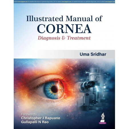
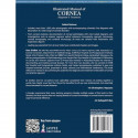
| Title | Illustrated Manual of Cornea: Diagnosis and Treatment for Ophthalmology Residents and Cornea Specialists | Corneal Disorders with 1000+ Color Photographs, Schematic Diagrams and Advanced Imaging Tech |
| Author | Uma Sridhar |
| ISBN-13 | 9789372026283 |
| Publication | Jaypee Brothers Medical Publishers |
 For General Enquiries E-mail support@madrasshoppe.com
 COD Available. Delivery Time 4 - 7 Working Days in India.
 Trusted by over 100K+ customers. Verified and Trusted Cashfree Online Payment
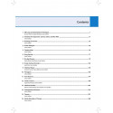
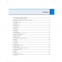
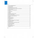
Illustrated Manual of Cornea: Diagnosis & Treatment, 1/E is a richly visual, scientifically thorough reference authored by Uma Sridhar with contributions from leading eye specialists across India. It features more than 1,000 high-quality color photographs paired with schematic line diagrams, offering clear documentation of a wide spectrum of corneal disorders. Structured across 31 chapters, the manual comprehensively covers corneal anatomy, documentation of corneal pathologies with color-coded diagrams, and advanced investigative techniques including anterior segment optical coherence tomography, specular microscopy, corneal tomography, and confocal microscopy. Every section pairs detailed illustrations with explanatory figures, ensuring clarity for learners working through corneal diagnosis for the first time as well as practitioners refining their diagnostic approach. Who This Book Is For: Postgraduate Ophthalmology Students: build a visual, case-referenced understanding of corneal anatomy and pathology for university and board examinations. Ophthalmology Residents: use the color-coded diagrams and photographs as a bedside and clinic reference for identifying corneal disorders. Ophthalmologists: refer to the imaging chapters (AS-OCT, specular and confocal microscopy) when working up complex corneal cases. Fellowship Trainees in Cornea and Anterior Segment: use the manual as a comprehensive, image-led resource through fellowship training. Why This Book Helps: Visual-first learning: more than 1,000 color photographs paired with schematic diagrams make pattern recognition faster and more reliable than text alone. Covers modern corneal imaging comprehensively: brings AS-OCT, specular microscopy, corneal tomography, and confocal microscopy together in one reference. Bridges anatomy to diagnosis: structures content from corneal anatomy through pathology documentation to investigation, mirroring real clinical workup. Multi-institutional expertise: chapters and images contributed by leading eye specialists from across India broaden the range of cases represented. Key Features: More than 1,000 high-quality color photographs with accompanying schematic line diagrams Color-coded diagrams documenting a wide range of corneal pathologies Dedicated coverage of anterior segment OCT, specular microscopy, corneal tomography, and confocal microscopy 31 chapters spanning corneal anatomy through advanced diagnostic investigation Contributions from leading eye specialists and cornea consultants across India
Uma Sridhar is the author of Illustrated Manual of Cornea: Diagnosis and Treatment for Ophthalmology Residents and Cornea Specialists | Corneal Disorders with 1000+ Color Photographs, Schematic Diagrams and Advanced Imaging Tech.
Disclaimer:
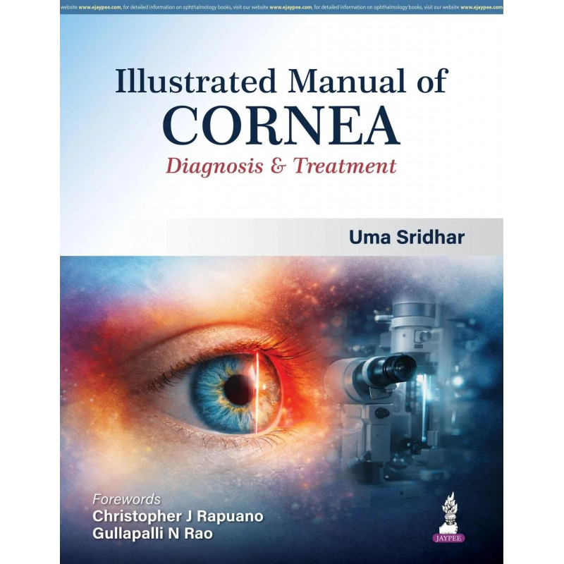
| Title | Illustrated Manual of Cornea: Diagnosis and Treatment for Ophthalmology Residents and Cornea Specialists | Corneal Disorders with 1000+ Color Photographs, Schematic Diagrams and Advanced Imaging Tech |
| Author | Uma Sridhar |
| ISBN-13 | 9789372026283 |
| Publication | Jaypee Brothers Medical Publishers |